
Enquire Now
About Us
Specialist in out patient Minimally invasive Neck & Back Herniated Disc Surgery. Dr Vangani has vast experience and he has one of the largest series of Minimally Invasive Herniated Disc Surgery in India.
FACT :- Not all Surgeons are alike, Innovations in Surgical Technology have enabled a handful of uniquely trained Surgeons to perform minimally disruptive surgery, resulting in less discomfort and faster recovery for the patient.
Dr Vangani has specialized in minimally invasive surgery for Herniated Cervical and Lumbar Discs. These unique out patient procedures frequently allows patient to feel relief immediately after the surgery is complete. The surgery usually last only 30 minutes.
After the surgery, majority of the Herniated Cervical disc patient relieved of their neck and arm pain and Lumbar Herniated disc patients are relieved of their back and leg pain. Using an operating microscope Dr Vangani also performs more traditional spinal operations such as anterior cervical discectomy for Cervical Spine Spondylosis and Lumbar Laminectomy for spinal stenosis.
Many of our patients are physically active and are involved in various sports activity. Dr Vangani is keenly aware of their need to return to their previous performance level and tries very hard to achieve it. Also because of Dr Vangani's reputation, many of his patients come from various fields of medicine i.e. physician, nurses, gynecologist, psychologist as well as policeman, fireman, bank employees etc.
Dr Vangani frequently presents lecture and live demonstration of procedure of Cervical and Lumbar disc at many places in India.

Dr Deepak vangani
MBBS, MS, MCH
Dr Deepak Vangani is a known spine surgeon who trained in Bombay
University presently working as consultant Neuro-spinal Surgeon in Jaipur (INDIA).
- - Brain tumor
- - Spine tumor
- - Minimally invasive surgery
- - Laser spine surgery
- - Microsurgery
- - Head Injury & Spine Injury
Specialist In
Information About Spine
The spinal column is one of the most vital parts of the human body, supporting our trunks and making all of our movements possible. When the spine is injured and its function is impaired the consequences can be painful and even disabling. According to estimates, 80 percent will experience low back pain at least once in their lifetime. A small number of patients will develop chronic or degenerative spinal disorders that can be disabling. Men and women are equally affected by lower back pain, and most back pain occurs between the ages of 25 and 60. However, no age is completely immune. Approximately 12% to 26% of children and adolescents suffer from low back pain. Fortunately most low back pain is acute, and will resolve itself in three days to six weeks with or without treatment. If pain and symptoms persist for longer than 3 months to a year, the condition is considered chronic.
The spinal column consists of 24 separate bones, called vertebrae, plus the fused bones of the sacrum and the coccyx. Your spinal column is the central support for the upper body, carrying most of the weight of your head, chest, and arms. Together with the muscles and ligaments of your back, your spinal column enables you to walk upright.
The Cervical region has seven vertebrae (C1 through C7), the Thoracic region has 12 vertebrae (T1 through T12) and the Lumbar region has five vertebrae (L1 through L5). The Sacral region consists of five vertebrae, all fused together to form one continuous bone mass known as the sacrum. The Coccygeal region consists of four vertebrae, all fused together to form the coccyx or tailbone.
Spine Parts & Its Importance
The Cervical Vertebrae
The vertebrae in the cervical (or upper back) portion of your spine carry the weight of your head. The pressure from this weight along with the "wear and tear" associated with the constant turning and bending of your head and neck is what usually leads to problems associated with the cervical vertebrae.
The Intervertebral Vertebrae
The intervertebral discs are composed of a fiber-like outer lining (the annulus) and a gelatin-like inner core (the nucleus). These discs act as the spine's "shock absorbers," preventing vertebra from rubbing against one another and providing much of the flexibility found in your spine. Because they are under constant pressure, it is the intervertebral discs which first show signs of the "wear and tear" associated with the aging process.
The Spinal Nerves
Running through the center of the spinal column is the spinal cord, which ends in the lumbar spine in a bundle of nerves called the cauda equina. At each disc level, a pair of spinal nerve roots branch off from the spinal cord or cauda equina and pass through an opening called the foramen.
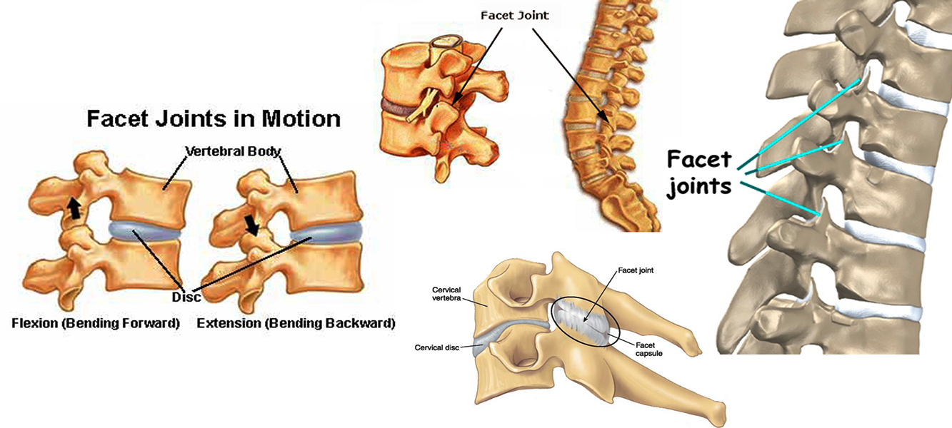
The Facet Joints
In the back of the spine hinge-like structures called facet joints are located. These joints link the vertebrae together along with the discs in front allowing for complex spinal motion. These joints may be injured by lifting or straining, severe twisting or hyperextension. Rest, ice and a few Aspirin will often help the pain. Facet injections with a small amount of anesthetic and cortisone are sometimes performed in resistant cases
The Growth of Bone Spurs
As your spine ages, the gelatin-like centers of your discs begin to dry out, thereby reducing their effectiveness as "shock absorbers". As this protection is lost, the simple "wear and tear" of everyday activity can cause the bone matter of your vertebrae to develop jagged edges, called bone spurs. As these spurs develop and extend outward, they can cause both the spinal canal and the foramen to become narrowed. The result is often the pinching (compression) of the spinal cord and/or a spinal nerve root.
The Slowly Closing Window
As discs dry out, your vertebrae begin to "settle." This "settling" causes the window-like openings of the foramen and the spinal canal to become smaller and smaller. Eventually, these openings can become so small that a spinal nerve(s) becomes "pinched" against a vertebra. It's similar to slowly closing a window on your hand. There will be a point at which you begin to feel the pressure. The more the window is closed, the greater the pressure and the greater the pain.
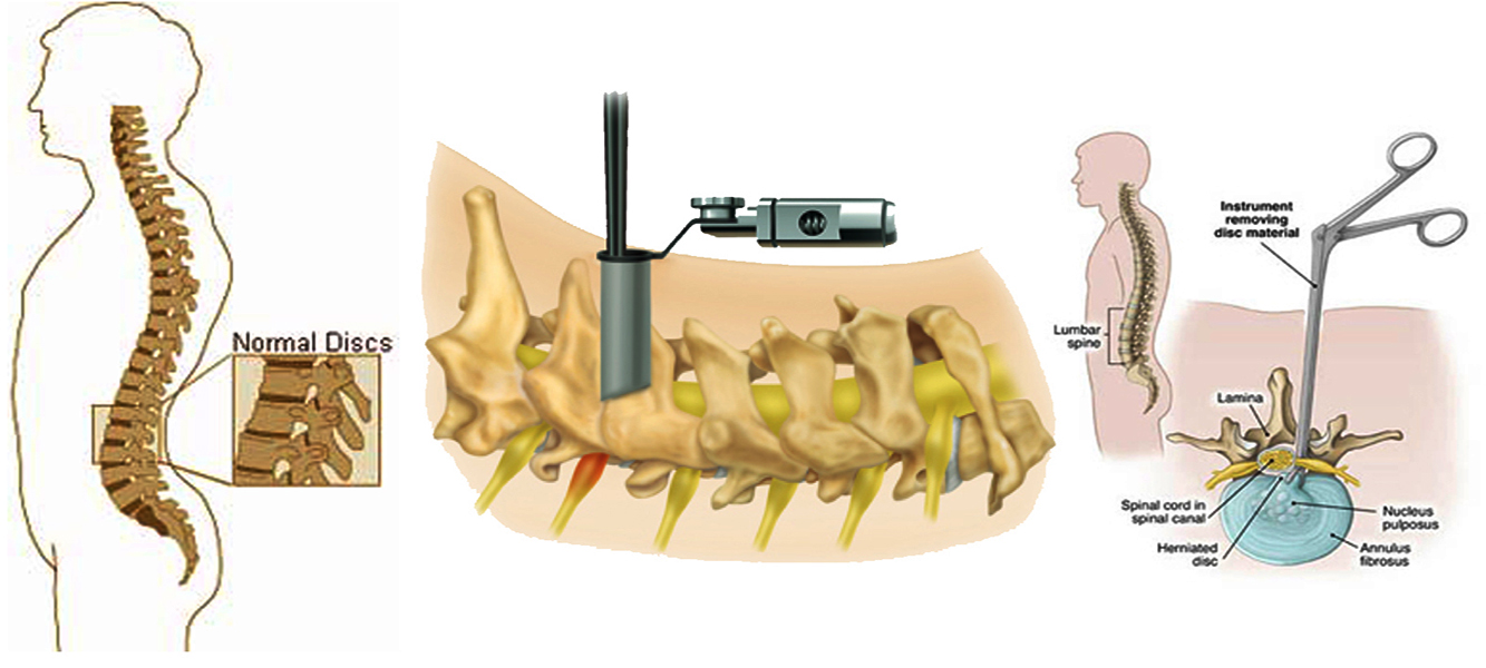
Endoscopic Discectomy
An Endoscopic Discectomy is the surgical removal of intervertebral disc with an endoscope (a device consisting of tube and Optical System for observing the inside of hollow organ or space). An endoscopic discectomy may be done through a small puncture opening.
Endoscopic Discectomy is an OPD surgical procedure to remove herniated disc material. Using local anesthesia with the help of XRay fluoroscopy and magnified video for guidance, a small specially designed endoscopic probe is inserted between the Vertebra and into Herniated disc space through skin of the back. Tiny Surgical attachments are then sent down the hollow center of the probe to remove a portion of the offending disc. Sometimes, the microsurgical attachments can be used to push the bulging disc back into place and be used for the removal of disc fragments and small burn spurs.
On average, the procedure takes about an hour. XRay exposure is minimal. You normally will feel little, if any pain or discomfort. Upon completion, the probe is removed and a small Band-Aid is placed over the incision. The amount of nucleus tissue removed varies and the supporting structure of the disc is not affected by surgery.
An Endoscopic Discectomy is different from an open disc surgery, because there is no traumatic muscle dissection, no bone removal and no large skin incision. The risk of complications from scarring, blood loss, infection and anesthesia that may occur with conventional surgery are drastically reduced and eliminated with this procedure. Endoscopic Discectomy was invented to be an effective treatment for herniated disc while avoiding these risks. Endoscopic discectomy is truely a minimally invasive procedure

NEURO SPINE CLINIC
L-24, Income Tax Colony, Durgapura Jaipur-302 018 (Rajasthan) India
For Appointment Call
+91-9829013398